
3D Paleontology
New technologies for studying fossils
COLLECTIONSRESEARCH
Blanca Moncunill-Solé
6/21/20232 min read
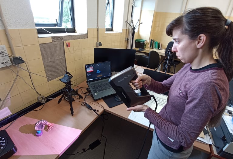
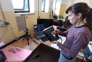
These last days, I have been practicing with a structured-light 3D scanner that we have in the Vertebrate Paleontology lab of GRICA. The light source of this kind of scanners projects a series of parallel patterns onto the target, in this case an ammonite reconstruction. The surface of the target distorts the pattern and the cameras captures these images. The software of the scanner processes this information, giving as a result a 3D model.
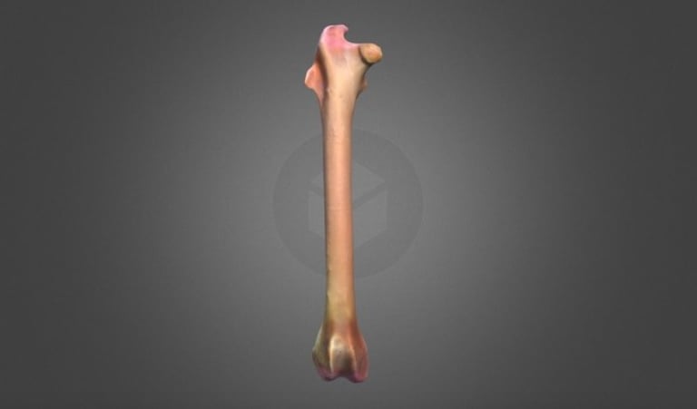
Virtual Paleontology emerges in recent decades, and it is described as the "study of fossils through 3D digital visualizations or virtual fossils". The construction of 3D models let us to extract data from the remains that in some cases cannot be gained from other approaches and are used for quantitative studies or functional morphology. In addition, virtual paleontologists can isolate parts of the specimens without damaging the real remain (e.g., cranial bones) or mark them in different colors (e.g., improving the visualization and clarity). In this regard, the researchers working on virtual paleontology do not require expensive lab-bound microscopes, and can research only using their laptop. The 3D models can be shared with other professionals in the field that want to work with this fossil remain (while the fossil continues curated in the collection without being damaged). These models can be used for outreach and educational purposes as well.
Several techniques exist to capture the shape of a fossil. Tomography (CT) let us to obtain images of the inner structure of the object, and the shape reconstruction of the element. On the other hand, there are several techniques to capture the surface of the object (shape), but not the inner structure. In this category, we can find laser scanning and mechanical digitization, but also the photogrammetry. This last technique, based on multiple 2D photographs, is able to reconstruct the surface topography of an object. In the past, I used CT and photogrammetry to obtain 3D models of lagomorphs fossils (see my Sketchfab profile).
Eager to continue practicing with this and other new techonologies!